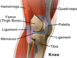
The knee is the largest joint and one of the most important joints in the body. It plays an essential role in movement related to carrying the body weight in horizontal (running and walking) and vertical (jumping) directions.
The knee is a joint that has three compartments. This joint has an inner (medial) and an outer (lateral) compartment. The kneecap (patella) joins the femur to form a third compartment called the patellofemoral joint. The thigh bone (femur) meets the large shinbone (tibia) forming the main knee joint. These ligaments provide stability and strength to the knee joint.
- Femur: The femur or thigh bone, is the most proximal (closest to the hip joint) bone of the leg in tetrapod vertebrates capable of walking or jumping. The head of the femur articulates with the acetabulum in the pelvic bone forming the hip joint, while the distal part of the femur articulates with the tibia and kneecap forming the knee joint. By most measures the femur is the strongest bone in the body. The femur is also the longest bone in the body.
- Patella: The patella , also known as the kneecap or kneepan, is a thick, circular-triangular bone which articulates with the femur (thigh bone) and covers and protects the anterior articular surface of the knee joint.
In humans, the patella is the largest sesamoid bone in the body. Babies are born with a patella of soft cartilage which begins to ossify into bone at about three years of age. - Lateral Collateral Ligament: The fibular collateral ligament (long external lateral ligament or lateral collateral ligament, LCL) is a ligament located on the lateral (outer) side of the knee, and thus belongs to the extrinsic knee ligaments and posterolateral corner of the knee.
- Medial Collateral Ligament: The medial collateral ligament (MCL), or tibial collateral ligament (TCL), is one of the four major ligaments of the knee. It is on the medial (inner) side of the knee joint in humans and other primates. Its primary function is to resist valgus forces on the knee.
- Lateral Meniscus: The lateral meniscus (external semilunar fibrocartilage) is a fibrocartilaginous band that spans the lateral side of the interior of the knee joint. It is one of two menisci of the knee, the other being the medial meniscus. It is nearly circular and covers a larger portion of the articular surface than the medial. It can occasionally be injured or torn by twisting the knee or applying direct force.
- Medial Meniscus: The medial meniscus is a fibrocartilage semicircular band that spans the knee joint medially, located between the medial condyle of the femur and the medial condyle of the tibia. It is also referred to as the internal semilunar fibrocartilage. The medial meniscus has more of a crescent shape while the lateral meniscus is more circular. The anterior aspects of both menisci are connected by the transverse ligament. It is a common site of injury, especially if the knee is twisted.
Was this article helpful?
YesNo