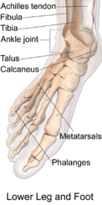
The ankle joint
The ankle joint is a synovial hinge joint, so you can plantarflex and dorsiflex. It allows a little wiggle from side to side, but most of the rest of the movement comes from the foot joints. The ankle joint is made up of distal ends of the tibia and fibula, which form a socket that fits over the top portion of the talus. The bones are held together by several ligaments.
- Medial (deltoid) ligament of the ankle: The medial malleolus. It has four parts named for the bones that they attach to: the tibionavicular, the tibiocalcaneal, the anterior tibiotalar, and the posterior tibiotalar.
- Lateral ligament of the ankle: This ligament is made up of three bands that start at the lateral malleolus. These ligaments are named for the bones they attach to:
1. Anterior talofibular ligament: This ligament runs to the lateral surface of the talus.
2. Calcaneofibular ligament: This ligament runs to the lateral surface of the calcaneus.
3. Posterior talofibular ligament: This ligament runs to the lateral tubercle (small rounded eminence) of the talus.
If you’ve ever sprained an ankle, you injured one or more of the ligaments that hold the joint together. The lateral ligaments are damaged more often than the stronger medial ligament.
The foot and toe joints
The foot contains a number of joints, but two important joints are the subtalar and transverse tarsal joints. These two joints allow you to invert and evert the foot.
- Subtalar joint:
In human anatomy, the subtalar joint, also known as the talocalcaneal joint, is a joint of the foot. It occurs at the meeting point of the talus and the calcaneus. - Transverse tarsal joint:
The transverse tarsal joint is actually a combination of the following two joints:
- Talocalcaneonavicular joint: The talocalcaneonavicular joint is a ball and socket joint: the rounded head of the talus being received into the concavity formed by the posterior surface of the navicular, the anterior articular surface of the calcaneus, and the upper surface of the plantar calcaneonavicular ligament.
- Calcaneocuboid joint: The calcaneocuboid joint is the joint between the calcaneus and the cuboid bone. The calcaneocuboid joint is conventionally described as among the least mobile joints in the human foot. The articular surfaces of the two bones are relatively flat with some irregular undulations, which seem to suggest movement limited to a single rotation and some translation. However, the cuboid rotates as much as 25 degree about an oblique axis during inversion-eversion in a movement that could be called obvolution-involution.
The remaining joints of the foot allow for a little movement of the foot and toes:
- Cuneonavicular joint: This synovial joint is formed between the navicular bone and the three cuneiform bones. It is supported by dorsal and plantar cuneonavicular ligaments. It allows for some gliding movement.
- Cuboideonavicular joint: This fibrous joint is between the cuboid and navicular bones. It’s supported by dorsal, plantar, and interosseous ligaments.
- Tarsometatarsal joints: These synovial joints are formed between the tarsal bones and the bases of the metatarsal bones. These joints are strengthened by dorsal, plantar, and interosseus ligaments.
- Intermetatarsal joints: These synovial joints involve the bases of the metatarsal bones. All these joints are strengthened by dorsal, plantar, and interosseus ligaments.
- Metatarsophalangeal joints: These synovial joints are between the heads of the metatarsal bones and the bases of the proximal phalanges. They’re supported by plantar and collateral ligaments. They allow you to flex and extend your toes as well as move them apart and closer together.
- Interphalangeal joints: These joints connect the phalanges. They’re synovial joints strengthened by collateral and plantar ligaments, and they let you flex and extend your toes.