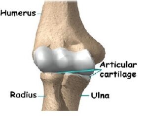
At first, the elbow seems like a simple hinge. But when the complexity of the interaction of the elbow with the forearm and wrist is understood, it is easy to see why the elbow can cause problems when it does not function correctly. Part of what makes us human is the way we are able to use our hands. Effective use of our hands requires stable, painless elbow joints.
- bones and joints
- ligaments and tendons
- muscles
- nerves
- blood vessels
The elbow joint has three different portions surrounded by a common joint capsule. These are joints between the three bones of the elbow, the humerus of the upper arm, and the radius and the ulna of the forearm.
When in anatomical position there are four main bony landmarks of the elbow. At the lower part of the humerus are the medial and lateral epicondyles, on the side closest to the body (medial) and on the side away from the body (lateral) surfaces. The third landmark is the olecranon found at the head of the ulna. These lie on a horizontal line called the Hueter line. When the elbow is flexed, they form an equilateral triangle called the Hueter triangle.
Humeroulnar joint: is part of the elbow-joint. It composed of two bones, the humerus and ulna, and is the junction between the trochlear notch of ulna and the trochlea of humerus. It is classified as a simple hinge-joint, which allows for movements of flexion, extension and circumduction. Owing to the obliquity of the trochlea of the humerus, this movement does not take place in the antero-posterior plane of the body of the humerus.
When the forearm is extended and supinated, the axis of the arm and forearm are not in the same line; the arm forms an obtuse angle with the forearm, known as the carrying angle. During flexion, however, the forearm and the hand tend to approach the middle line of the body, and thus enable the hand to be easily carried to the face.
The accurate adaptation of the trochlea of the humerus, with its prominences and depressions, to the trochlear notch of the ulna, prevents any lateral movement.
Flexion in the humeroulnar joint is produced by the action of the biceps brachii and brachialis, assisted by the brachioradialis, with a tiny contribution from the muscles arising from the medial epicondyle of the humerus.
Extension in the humeroulnar joint is produced by the triceps brachii and anconeus muscle, with a tiny contribution from the muscles arising from the lateral epicondyle of the humerus, such as the extensor digitorum muscle.
Humeroradial joint: The humeroradial joint is the joint between the head of the radius and the capitulum of the humerus, is a limited ball-and-socket joint, hinge type of synovial joint.
The bony surfaces would of themselves constitute an enarthrosis and allow movement in all directions, were it not for the annular ligament, by which the head of the radius is bound to the radial notch of the ulna, and which prevents any separation of the two bones laterally.
The annular ligament secures the head of the radius from dislocation, which would otherwise tend to occur, from the shallowness of the cup-like surface on the head of the radius. Without this ligament, the tendon of the biceps brachii would be liable to pull the head of the radius out of the joint.
The head of the radius is not in complete contact with the capitulum of the humerus in all positions of the joint.
The capitulum occupies only the anterior and inferior surfaces of the lower end of the humerus, so that in complete extension a part of the radial head can be plainly felt projecting at the back of the joint.
In full flexion the movement of the radial head is hampered by the compression of the surrounding soft parts, so that the freest rotatory movement of the radius on the humerus (pronation and supination) takes place in semiflexion, in which position the two articular surfaces are in most intimate contact.
Flexion and extension of the elbow-joint are limited by the tension of the structures on the front and back of the joint; the limitation of flexion is also aided by the soft structures of the arm and forearm coming into contact.
Superior radioulnar joint:The proximal radioulnar articulation (superior radioulnar joint) is a synovial pivot joint between the circumference of the head of the radius and the ring formed by the radial notch of the ulna and the annular ligament.